

Illustrative stock photograph; not a CEI patient, treatment result or clinic equipment.
If you have glaucoma or are being watched for it, you’ve probably spent time looking into machines at your appointments. Two tests come up again and again: OCT scans and visual field tests. They can feel repetitive, but they’re two of the most important tools we have for protecting your sight.
Glaucoma damages the optic nerve slowly and usually without symptoms. By the time you notice a problem, a lot of nerve fibres may already be lost. These tests help us find changes early, long before you would, and decide whether your treatment is doing its job. Here’s what each one shows and how the results come together into a plan.
Key Takeaways
- Glaucoma slowly damages the optic nerve, usually with no early symptoms. Regular testing is how we catch changes.
- OCT is a quick, painless scan that measures the thickness of the nerve fibre layer and the optic nerve.
- A visual field test maps your side vision to show how well the optic nerve is working.
- The real value comes from comparing results over time. Analysis software helps us spot even small changes.
- Your results guide decisions about drops, laser or surgery, and how often you need to be seen.
Two Tests, Two Questions
Glaucoma testing answers two questions:
- What does the optic nerve look like? That’s what OCT shows. It looks at the structure of the nerve.
- How well is the optic nerve working? That’s what a visual field test shows. It measures function, how well you can see across your field of vision.
Using both gives a far more complete picture than either one alone. Along with your eye pressure measurements and a look at the optic nerve during your exam, they form the foundation of glaucoma care. Our post on glaucoma tests covers the full set.
OCT: A Detailed Look at the Optic Nerve
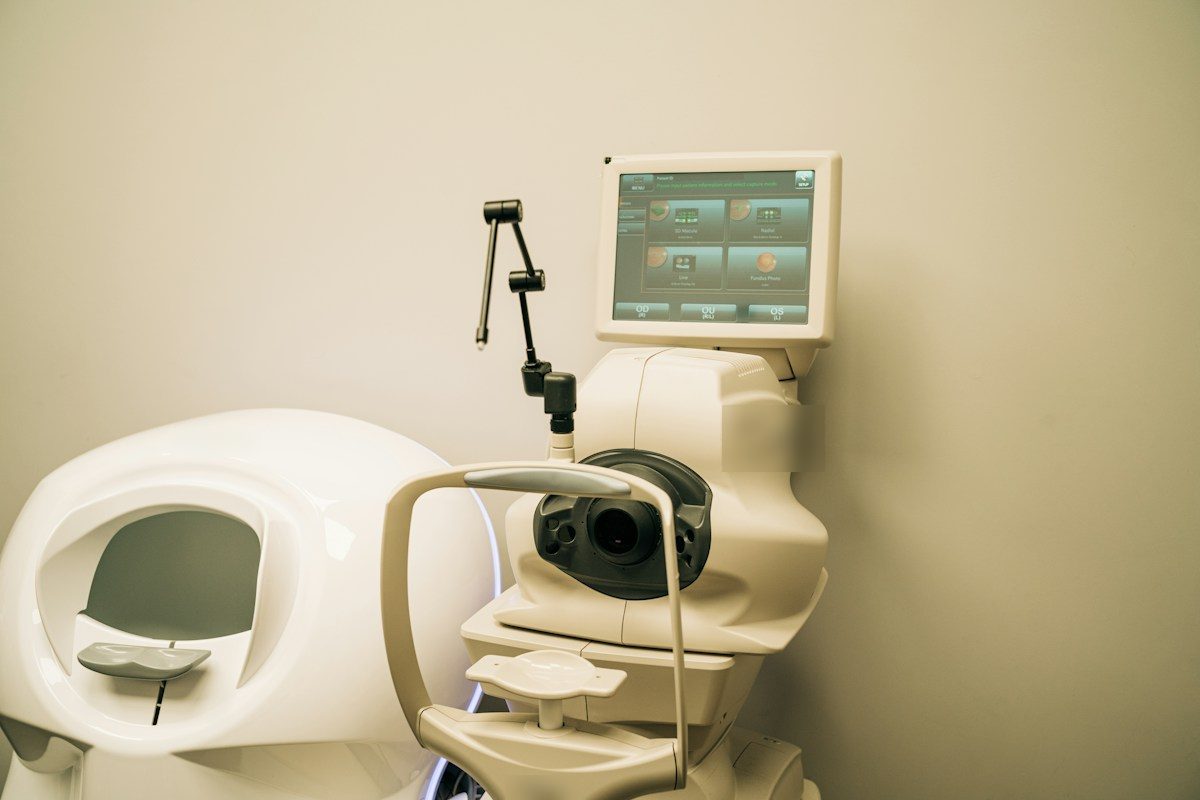
Optical coherence tomography (OCT) uses light to create detailed, cross-section images of the back of the eye. For glaucoma, we focus on:
- The retinal nerve fibre layer, the layer of nerve fibres that come together to form the optic nerve. Glaucoma thins this layer.
- The optic nerve head, where the nerve leaves the eye. We look at its shape and the rim of healthy tissue.
- The macula, where certain nerve cells affected by glaucoma are concentrated.
What to Expect
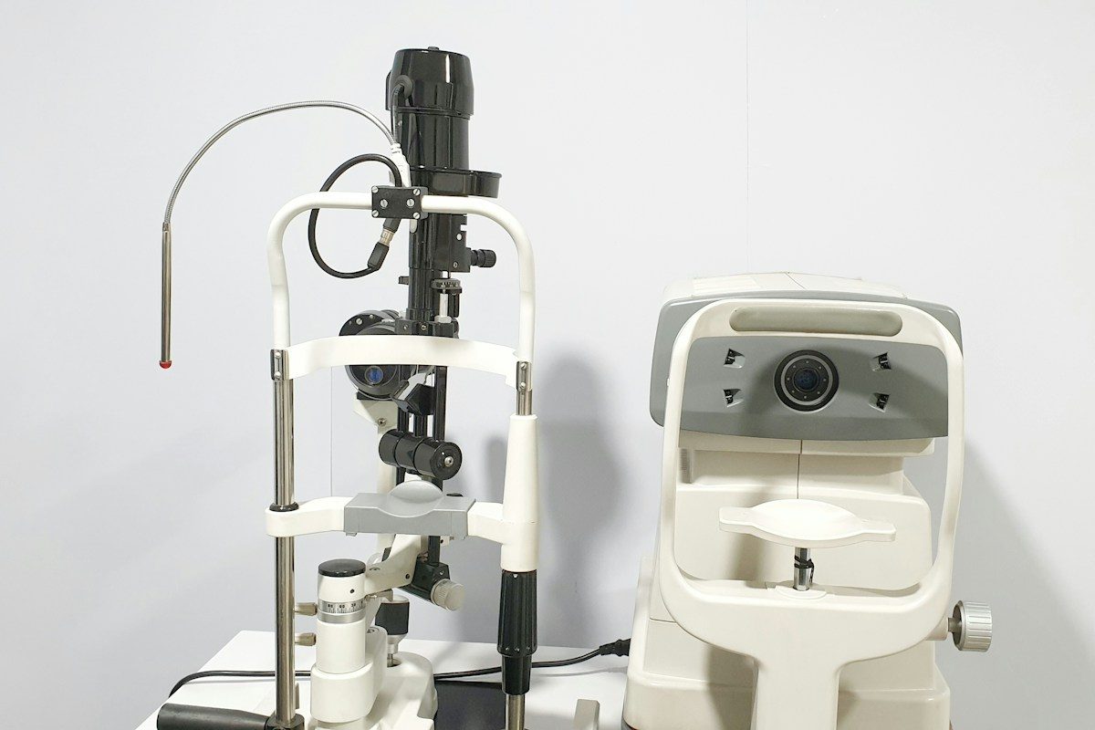
An OCT scan takes only a few minutes. You rest your chin on a support, look at a target light, and the machine scans your eye. Nothing touches your eye, and there’s no flash or discomfort. Sometimes we use dilating drops first.
Visual Field Testing: Mapping Your Side Vision
Glaucoma usually affects side vision first. Because the other eye and your brain fill in the gaps, you may not notice any loss until it’s advanced. A visual field test maps your vision to find these blind spots early.
What to Expect
You sit in front of a bowl-shaped instrument, cover one eye and look straight ahead at a central light. Small lights flash in different spots, some bright and some dim. You press a button each time you see one. Each eye takes several minutes.
A few tips:
- Missing some lights is normal. The test is designed to find the dimmest light you can see, so everyone misses some.
- Keep looking at the centre light. Don’t search for the flashes.
- Blink normally, and ask for a break if you need one.
- Wear the lens they give you if a lens is used to correct your focus.
- Get a good night’s sleep before. Being tired can affect your results.
Why We Repeat the Tests
A single OCT scan or visual field test tells us where things stand today. The real power is in comparing results over time.
Glaucoma care is about rate of change. If your tests are stable year after year, your treatment is working. If they show steady thinning or new blind spots, your treatment may need to be stepped up. Repeating tests also helps us separate a true change from an “off day.”
Glaucoma analysis software brings your OCT and visual field results together, lines them up over years and highlights changes that might be too small to spot by eye. That helps us make decisions sooner and with more confidence.
How Results Shape Your Plan
Your test results, along with your eye pressure, your age, your family history and your overall health, guide decisions such as:
- Whether to start treatment if you’re being watched for glaucoma or high eye pressure
- Whether your target pressure is right, or needs to be lower
- Whether to add or change drops, or consider laser treatment such as selective laser trabeculoplasty (SLT), which Conlon Eye Institute offers in Saskatoon, or surgery
- How often to see you. Stable patients may need fewer visits. Changing patients may need more.
Frequently Asked Questions
How often do I need OCT and visual field tests?
It depends on your type of glaucoma, how stable it is and your risk of progression. Many people are tested once or twice a year. Some need testing more often.
Do these tests hurt?
No. Neither test touches your eye or causes discomfort. If your pupils are dilated, your vision may be blurry and light-sensitive for a few hours.
Can these tests diagnose glaucoma on their own?
They’re important, but the diagnosis is based on your whole exam, including eye pressure, the appearance of the optic nerve and your history.
Can glaucoma damage be reversed?
No. That’s why early detection and regular monitoring matter so much. Treatment aims to prevent further damage.
Book Your Consultation
If you have questions about your glaucoma testing or treatment, our ophthalmologists can help. Book your consultation: call our Saskatoon office at 306-244-4111, Monday to Friday, 7 am to 4 pm. Conlon Eye Institute is in the Medical Arts Building, 341-750 Spadina Crescent East, Saskatoon, SK S7K 3H3.
References
- American Academy of Ophthalmology. Glaucoma Diagnosis. https://www.aao.org/eye-health/diseases/glaucoma-diagnosis — supports OCT, visual field testing, pressure and other tests used together to diagnose and monitor glaucoma.
- National Eye Institute. Glaucoma. https://www.nei.nih.gov/learn-about-eye-health/eye-conditions-and-diseases/glaucoma — supports that damage can’t be reversed, early detection and treatment with drops, laser or surgery.
About the Author
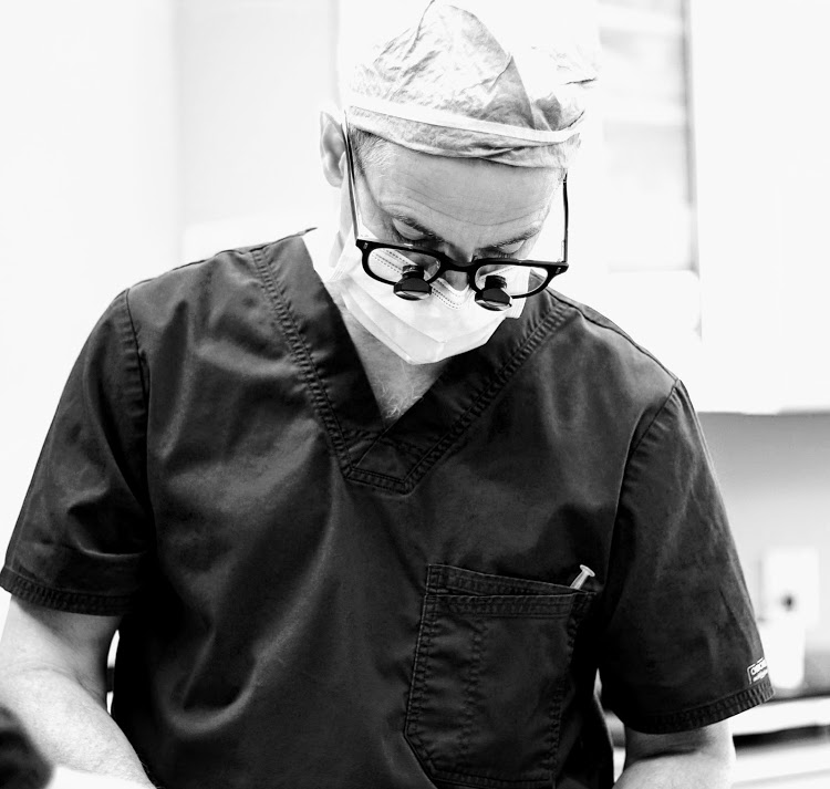
Dr. M. Ronan Conlon has practised ophthalmology in Saskatoon since 1994. He completed fellowship training in ocular pathology at Harvard University and in oculoplastic surgery at the University of Iowa. In 1996 he brought laser vision correction technology to Canada from Germany, and he specializes in oculoplastic surgery, LASIK, ASL (PRK) and refractive cataract surgery. With the establishment of the Conlon Eye Institute, Dr. Conlon has drawn on decades of experience in LASIK, RLE surgery, refractive cataract surgery, and oculoplastic surgery. He has also served as Head of Ophthalmology and Chief of Staff with the Saskatchewan Health Authority.
The information on this page should not be used in place of information provided by a doctor or specialist. To learn more, read our Privacy Policy and Editorial Policy pages.